
Lipomyelomeningocele and tethered cord Radiology Case
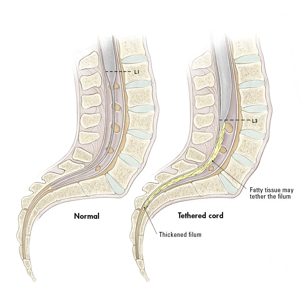
Tethered cord syndrome is defined as a stretch-induced clinical constellation arising from tension on the spinal cord due to caudal anchoring to inelastic structures. Inelastic structures restrict vertical movement of the spinal cord and may arise from congenital etiologies, such as myelomeningocele, or acquired etiologies, such as scar formation.

Dr. Arun L.Naik Adult Tethered Cord Syndrome
Neurosurgeon Deb Bhowmick, MD, who specializes in the technique, explains that surgery to release a tethered cord is effective, but the cord can retether, and the surgery can be performed a very limited number of times due to the high risk of injury. "So the patient is left with only pain relief, as they slowly lose the ability to walk," he.

Tethered cord Radiology Case
Tethered cord Low conus medullaris Thickened filum terminale Hydromyelia Spinal lipoma Dorsal dermal sinus Diastematomyelia Blunt cord terminus Classification of Spinal dysraphism Spina bifida aperta Spinal dysraphism or spina bifida is a congenital anomaly resulting in a defective closure of the neural arch.
TetheredCord
Tethered cord syndrome (TCS) has been well described in pediatric patients. Many recent reports of TCS in adult patients have grouped retethering patients with newly diagnosed ones without separately analyzing each entity and outcome.

Tethered Cord Kids health, Spinal cord, Chiari malformation
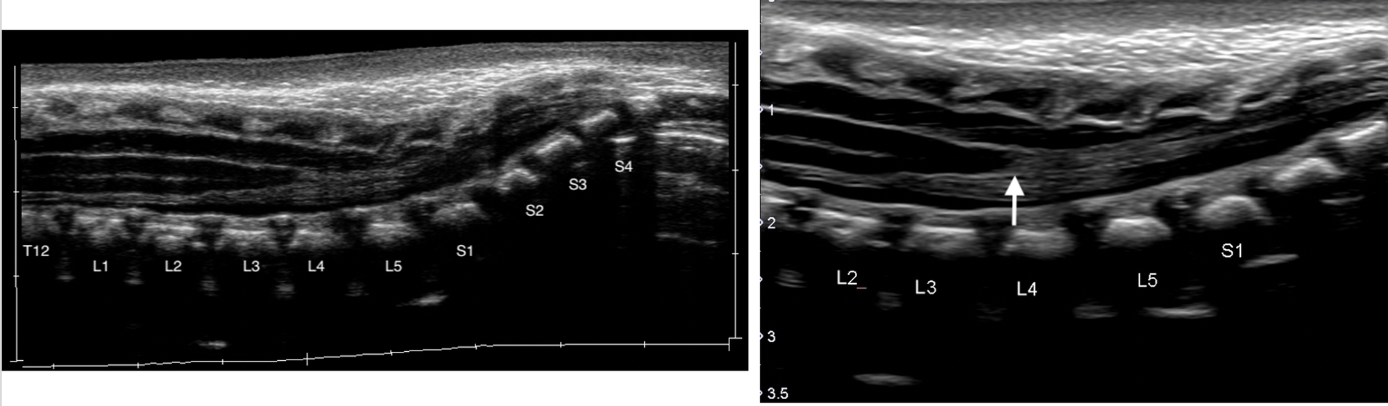
Tethered cord Case contributed by Maulik S Patel Diagnosis certain Share Add to Citation, DOI, disclosures and case data Presentation Club foot Patient Data Age: Neonate ultrasound Cord tethering at L1-L2 level. Lower end of cord at sacral level. Two small syringes in dorsal cord. No hydrocephalus. 1 article features images from this case
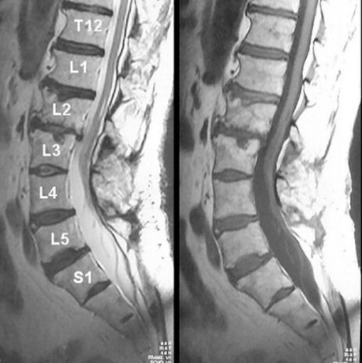
Cureus Tethered Cord Syndrome Associated With Lumbar Lipomyelomeningocele A Case Report
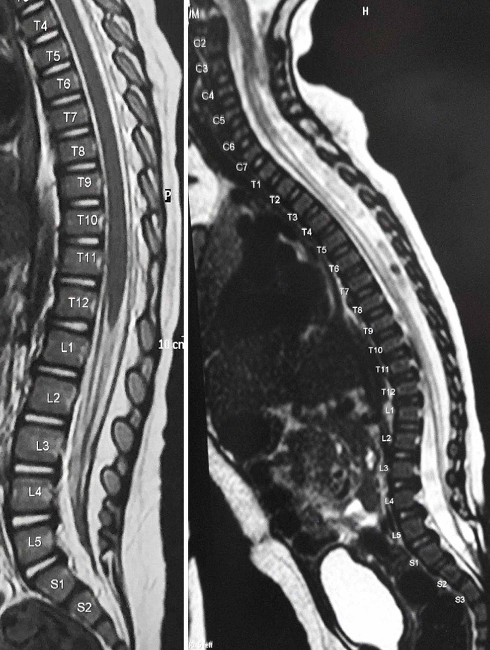
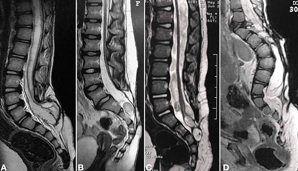
T1 Sagittal T2 3D Axial T2 Axial T2 S3 spina bifida with an open posterior neural arch that shows dorsal dermal sinus tract harboring dysplastic neurogenic and fibrous tissues. Associated superficially located subcutaneous loculus of CSF like intensities averaging 1 cm in size likely small meningocele.

Lspine MRI, Spinal Bifida with Tethered Cord YouTube
Confusingly, "tethered cord syndrome" is sometimes used synonymously, as if to imply that a tight filum terminale is the main etiology of the tethered cord syndrome, even though in actuality there are many other etiologies.
Cureus Tethered Cord Syndrome After Myelomeningocele Repair A Literature Update
T1 Sagittal T2 Axial T2 Axial T2 The vertebral bodies have normal alignment, height, and bone marrow signal. Incomplete posterior fusion at L5 and S1. Widening of the bony spinal canal from L3/4 to S1/2 due to dural ectasia. No spinal canal stenosis or cord compression.
Tethered Cord Syndrome
Introduction. The diagnosis of tethered cord syndrome (TCS) can be challenging to establish [], and the surgical solution is not without risk and does not always guarantee clinical improvement [].Prior studies have shown that standard magnetic resonance imaging (MRI) criteria such as a low-lying conus terminalis, or the presence of a fatty filum have a low imaging sensitivity and specificity.
Cureus Tethered Cord Syndrome After Myelomeningocele Repair A Literature Update
Tethered spinal cord syndrome occurs when surrounding tissue attaches to and causes stretching across the spinal cord. People with a tethered cord can experience weakness, pain, and loss of.

Image
Tethered spinal cord syndrome is a neurologic disorder caused by tissue attachments that limit the movement of the spinal cord within the spinal column. These attachments cause an abnormal stretching of the spinal cord. This syndrome is closely associated with spina bifida. It is estimated that 20-50% of children with spina bifida defects that.

Tethered cord Radiology Case
The tethered cord syndrome (TCS), also known as tight filum terminale syndrome is a clinical entity by which signs and symptoms are caused by excessive tension on the spinal cord. The majority of cases of tethered cord are related to spinal dysraphism.
Tethered Spinal Cord Syndrome Captions Save
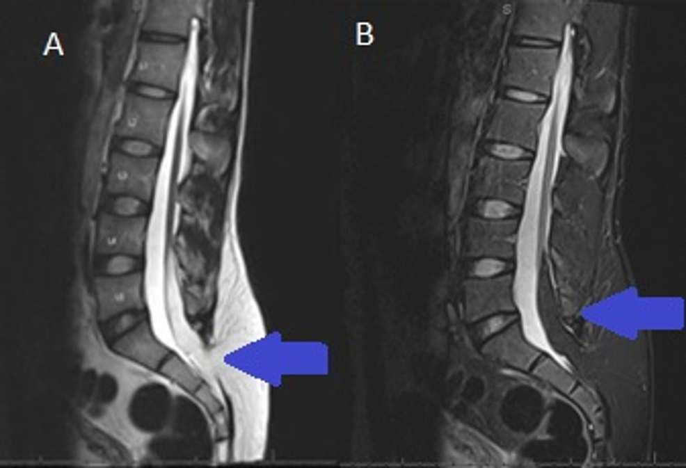
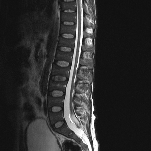
Classically, a tethered cord presents with symptoms including lumbosacral pain, lower extremity weakness, and neurogenic bladder dysfunction, as well as imaging findings of a low-lying, dorsally positioned conus medullaris with limited dependent movement [ 2 ].
Cureus A Novel Case of Tethered Cord in a FiveMonthOld Male With PallisterKillian Syndrome
Radiographic studies are used to confirm the presence of tethered cord, to ascertain the cause of tethering, and to rule out other diagnostic considerations such as neoplasms, disk herniations, and syringohydromyelia.
Tethered Spinal Cord Sumer's Radiology Blog
Tethered cord syndrome (TCS) is a clinical condition of various origins that arises from tension on the spinal cord.

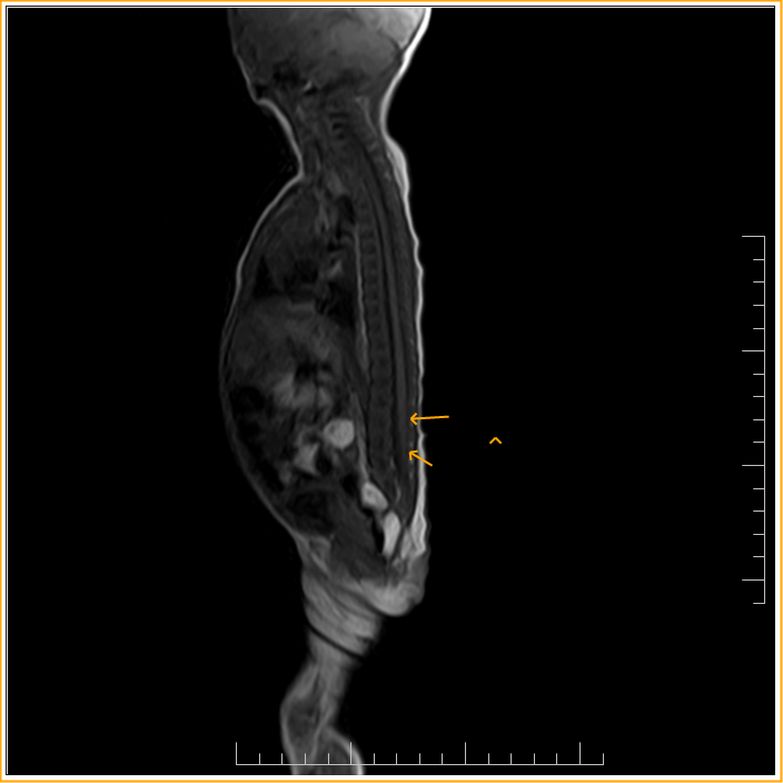
Tethered cord with terminal lipoma Radiology Case
Tethered cord syndrome is a neurological disorder caused by tissue attachments that limit the movement of the spinal cord within the spinal canal. Clinical presentation Tethered cord syndrome is a clinical diagnosis based on neurologic deterioration involving the lower spinal cord 7. Patients may present with any combination of the following 4: